
When retinitis pigmentosa takes a person’s sight, the standard clinical test goes flat: no response, no signal, nothing. Yet the retina behind that flat line is anything but quiet.
Researchers at the Jules-Gonin Eye Hospital, part of the Faculty of Biology and Medicine of the University of Lausanne, have recorded the spontaneous electrical activity of the retina—in darkness, with no light stimulation—and found it producing abnormal rhythms never before measured in a living person. The findings are published in Nature Communications.
Where today’s exams fall silent, the retina keeps talking
Retinitis pigmentosa is a group of genetic diseases that progressively destroy the photoreceptors, the cells that capture light. At an advanced stage, the electroretinogram—the reference test for retinal function—detects nothing. The trace is flat.
But a flat trace is not a dead retina. Behind the lost photoreceptors, several layers of neurons survive and rewire, a process called retinal remodeling that has been documented for some 20 years in donor tissue examined after death. What no one had done was measure the activity of those rewired circuits in a living eye.
A trick borrowed from the brain
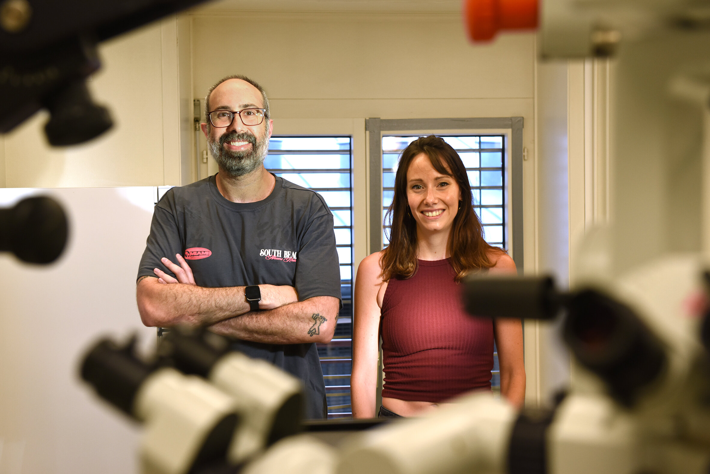
The team at the Ophthalmic and Neural Technologies Laboratory, led by Diego Ghezzi, Ph.D., adapted a principle from neurology. Resting-state EEG reads the brain’s spontaneous activity—no task, no stimulation—and reveals things that task-based tests miss.
Applied to the eye, that means recording the retina in the dark, with no flash and no image, and listening to the signal it generates on its own. The method is called resting-state electroretinography, or rsERG.
From mouse to patient
The first recordings used a flexible sensor placed on the cornea across three mouse models of retinitis pigmentosa. Every degenerating retina produced an oscillatory rhythm that was absent in healthy controls—detectable from the earliest stages of the disease and growing stronger as it advanced.
A pilot study then enrolled five patients at an advanced stage of the disease and eight sighted volunteers. Recordings lasted 40 minutes, eyes closed, in darkness, using thread electrodes already common in routine clinical care.
The patients’ retinas showed markedly stronger oscillatory activity than the sighted participants’, in a frequency range close to the one seen in mice—the first evidence that degenerating human retinas are functionally, not just anatomically, remodeled.
The noise can be turned down, and that helps
In animals, the team went a step further. A drug that boosts the retina’s own inhibitory signaling quieted the abnormal rhythm. And with the noise reduced, transcorneal electrical stimulation reached the visual cortex more effectively, at lower current, and the retina became easier to drive.
Why it matters
Therapies that aim to restore sight—retinal implants, optogenetics, regenerative approaches—all depend on the surviving retina being able to listen. If it is busy talking to itself, that is a problem. rsERG offers a way to measure that “oscillation burden” repeatedly and noninvasively as the disease progresses: to follow degeneration where current tests read nothing, to help identify who is ready for an implant or a restoration therapy, and to objectively gauge whether a treatment is working in a clinical trial.
The principle is not limited to retinitis pigmentosa and could extend to other conditions involving retinal remodeling, including age-related macular degeneration.
The authors are deliberately measured about what comes next. “This is a measurement, not a treatment,” says David Litvin, Ph.D., first author of the study. “It doesn’t restore vision, and it doesn’t change anyone’s prognosis today. We’re talking about years of validation, not something ready for the clinic. What this study does is make the direction credible and give us a noninvasive way to start.”
Publication details
David G. Litvin et al, Resting-state electroretinography reveals pathological retinal oscillations in retinitis pigmentosa mice and patients, Nature Communications (2026). DOI: 10.1038/s41467-026-75520-9
Journal information:
Nature Communications
Key medical concepts
Clinical categories


Citation:
A blind retina isn’t silent—and its noise may hinder efforts to restore sight (2026, September 15)
retrieved 17 October 2026
from https://medicalxpress.com/news/2026-09-retina-isnt-silent-noise-hinder.html
This document is subject to copyright. Apart from any fair dealing for the purpose of private study or research, no
part may be reproduced without the written permission. The content is provided for information purposes only.