
CT images taken before patients begin treatment with bone resorption inhibitors may contain measurable clues about who will later develop medication-related osteonecrosis of the jaw (MRONJ), according to a retrospective study by a researcher at Hiroshima University.
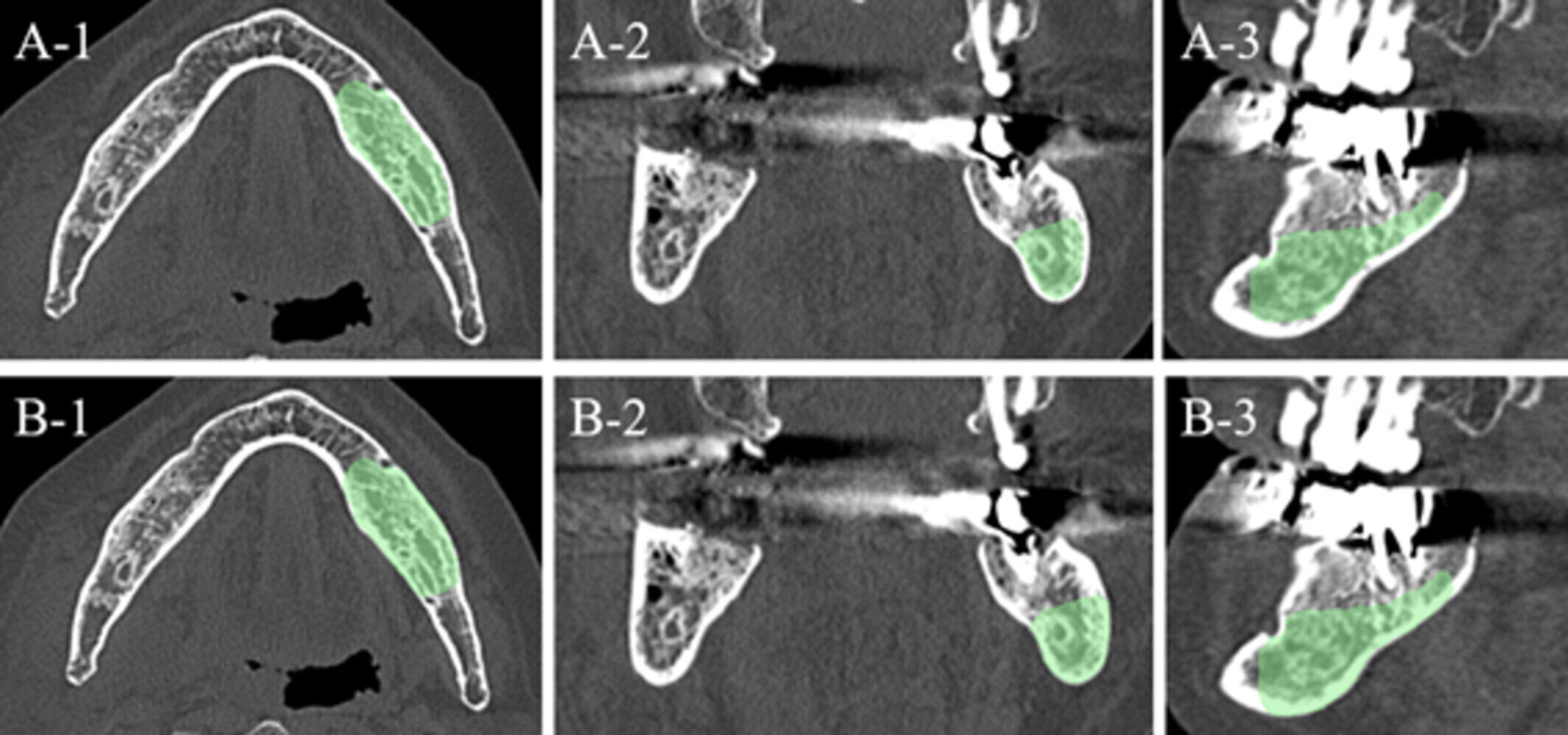
Masaru Konishi, a lecturer at Hiroshima University Hospital and sole author of the study published in Oral Diseases, used radiomics, which turns subtle patterns such as shape, texture and intensity in medical images into measurable data, to analyze the mandibular region in pretreatment CT scans.
When these measurements were combined with machine learning, the models distinguished patients who later developed MRONJ from those who remained unaffected. Analyses that included both the jaw’s porous, honeycomb-like cancellous bone and its denser, stronger cortical bone generally produced better predictive results than analyses limited to cancellous bone alone.
The stakes of early detection
MRONJ is a serious condition in which jawbone tissue dies and becomes exposed. It most often affects patients receiving bone resorption inhibitors, medications commonly used to strengthen bones in people with osteoporosis or bone metastases from cancer. Although MRONJ is relatively uncommon, some studies have reported incidence rates of up to 15% among patients receiving bone resorption inhibitors.
Once the condition develops, it can be difficult to treat without surgical intervention. In severe cases, treatment may require removal of part or all of the lower jawbone, which can lead to facial changes, difficulty chewing and biting, and numbness caused by nerve damage.
Intervention before MRONJ develops is therefore crucial to avoid life-altering surgeries and their complications. However, there is currently no established way to determine before treatment which patients are most likely to develop the condition. Identifying patients at higher risk before treatment begins could eventually support closer monitoring and more individualized treatment decisions.
Cortical bone sharpened predictions
The study analyzed pretreatment multidetector row computed tomography (MDCT) scans from 94 patients: 44 who later developed MRONJ in the lower jaw and 50 who remained free of the condition for at least two years after beginning bone resorption inhibitor therapy. Radiomic measurements were extracted from two regions: cancellous bone alone and a combined region containing both cancellous and cortical bone.
The machine-learning models generally performed substantially better when cortical bone was included. When the analysis was expanded from the jaw’s cancellous bone alone to include both cancellous and cortical bone, the Random Forest model’s average area under the curve (AUC)—a measure of how well a model distinguishes between patients who develop a condition and those who do not—increased from 0.741 to 0.931. The Support Vector Machine (SVM) model improved from 0.706 to 0.917, while the Multilayer Perceptron (MLP) model rose from 0.802 to 0.921.
An AUC of 1.0 represents perfect discrimination, while 0.5 indicates performance no better than chance. The results therefore suggest strong discrimination within the study population. In one analysis of the combined bone region, the MLP model correctly identified 88.6% of patients who later developed MRONJ and 88.6% of those who did not.
“This study demonstrated the potential to predict the development of MRONJ using image features extracted from CT images acquired prior to the administration of bone resorption inhibitors,” said Konishi.
Limits now, screening later
The CT images and machine-learning technologies show promise for accurately predicting mandibular MRONJ occurrence, though the study is not without limitations. For instance, the retrospective nature of the study does not necessarily guarantee consistent standards across the board. Patients’ dental factors also appear to be crucial to the development of MRONJ but were not necessarily captured in hindsight. Additionally, machine learning identifies potential relationships between features in the images and MRONJ occurrence, but the exact characteristics involved are not known.
Future work involves determining the defining characteristics identified in the images, such as bone quality or microarchitecture, as well as patients’ dental health information before the onset of MRONJ. Ultimately, Konishi would like to see the development of software capable of accurately predicting MRONJ occurrence based on radiographic images to help prevent the condition’s onset, allowing doctors to alter treatment plans and, if necessary, avoid prescribing bone resorption inhibitors or seek other treatment methods for patients who appear to have a high likelihood of developing MRONJ.
More information
Masaru Konishi, Radiomics Analysis for Predicting Medication‐Related Osteonecrosis of the Jaw Using Computed Tomography, Oral Diseases (2026). DOI: 10.1111/odi.70386
Key medical concepts
Clinical categories


Citation:
Pretreatment CT scans may contain clues about the risk of medication-related osteonecrosis of the jaw (2026, August 7)
retrieved 17 October 2026
from https://medicalxpress.com/news/2026-08-pretreatment-ct-scans-clues-medication.html
This document is subject to copyright. Apart from any fair dealing for the purpose of private study or research, no
part may be reproduced without the written permission. The content is provided for information purposes only.