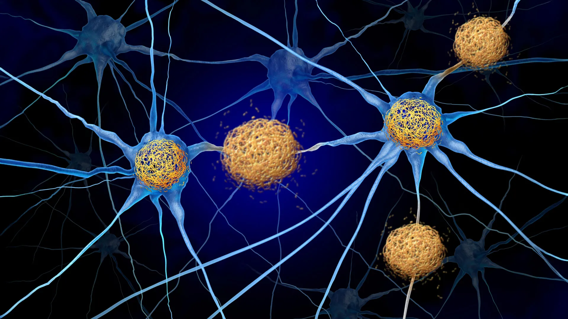
Folate-based radiopharmaceuticals can be used in positron emission tomography (PET) imaging to detect folate receptors in brain tumours. The discovery of folate receptors and their exploitation potential with respect to brain tumours is a new and significant finding in the field.
The discovery is related to gliomas, which are a group of serious brain tumours. Researchers discovered that brain tumours contain increased amount of folate receptor expression relative to adjacent brain tissue. This phenomenon has been observed in both experimental models and human tumour samples.
“Prior to this discovery, the presence of folate receptors and their increased presence in gliomas had not been recognised, and thus they have not yet been used for imaging nor treatment purposes,” summarises Doctoral Researcher Maxwell Miner from the Turku PET Centre at the University of Turku in Finland.
According to research group leader and InFLAMES PI Professor Anne Roivainen this presents an especially exciting target for potential future treatments.
“Our results show an average of 100-fold increase in folate-based radiopharmaceutical accumulation in glioma tissue versus that of adjacent healthy brain tissue,” says Professor Roivainen.
Urgent need for new chemotherapy treatments
Glioma brain tumours originate from the non-neuronal glial cells in the brain, which outnumber neurons in quantity. Gliomas comprise numerous subgroups, with even a high degree of morphological and receptor variability within a single cancerous lesion.
This exceptional cellular heterogeneity can make treatment difficult. There is an urgent need for new chemotherapy treatments particularly for the most malignant brain cancers as they often grow in an infiltrative web-like manner on their periphery making distinguishing the boundaries between glioma and non-glioma difficult. The researchers at the Turku PET Centre hope that this recent discovery will lead to further investigation into folate-targeted brain tumour detection and treatment.
The results were obtained in a multidisciplinary joint project involving researchers from the Turku PET Centre at the University of Turku, Turku University Hospital, InFLAMES Research Flagship, and collaborators from Purdue University, USA. The glioma samples were obtained from the Auria Biobank.