
In the United States, age-related macular degeneration is a leading cause of irreversible vision loss in people who are 60 and older.
It affects the central portion of the retina, called the macula. This region is packed with cells responsible for high-resolution color vision.
About 20 million U.S. adults are living with some form of AMD. Although they cannot see objects that are directly in front of them, their peripheral vision is unaffected.
Current treatments slow the disease, but none restore vision.
In a study, published in Cell Stem Cell, researchers used retinal pigment epithelial stem cells derived from adult postmortem eye tissue in a phase 1/2a clinical trial. These early phase trials are used to determine whether a therapy intervention is safe.
There are two types of macular degeneration: dry and wet.
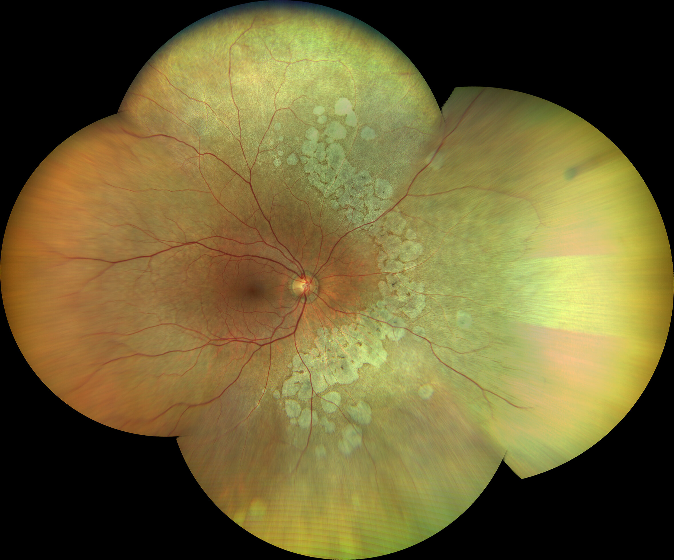
More than 90% of people with this condition have the dry form, which is caused by dysfunction and eventual loss of retinal pigment epithelial cells.
In early stages of AMD, these cells do not function properly. In late stages, they die and do not regenerate.
As the disease progresses, several areas inside the central eye lose these cells.
In the current study, patients with advanced dry AMD received transplanted stem cells, originally isolated from eye-bank tissues. These adult stem cells were specialized and could only develop into retinal pigment epithelial cells.
Six patients received the lowest dose of transplanted stem cells (50,000 cells) through a surgical eye procedure.
In all of them, the treatment was safe and did not cause serious inflammation or tumor formation.
The participants also experienced improved vision in the transplanted eye; the non-transplanted eye did not have these improvements, hinting that the approach could provide a new therapeutic avenue. “Although we were pleased with the safety data, the exciting part was that their vision was also improving,” said Rajesh C. Rao, M.D., Leonard G. Miller Professor of Ophthalmology & Visual Sciences, and an associate professor of pathology and human genetics.
“We were surprised by the magnitude of vision gain in the most severely affected patients who received the adult stem cell-derived RPE transplants. This level of vision gain has not been seen in this group of patients with advanced dry AMD.”
When tested with an eye chart, the participants in the low-dose group were able to see 21 more letters after a year.
The team is now following the 12 other patients who received medium and high doses of 150,000 and 250,000 cells.
If no safety concerns arise, the research team will proceed with the next phases of the clinical trial.
“We are grateful to all our participants who are allowing to better understand whether this intervention is safe enough to be a future therapy,” Rao said.
“These kinds of studies can help us offer advanced treatments in the field of regenerative medicine, and we are happy we can offer this first-in-human, cutting-edge clinical trial at the University of Michigan.”
More information:
Rajesh C. Rao et al, Safety and tolerability of RPESC-RPE transplantation in patients with dry age-related macular degeneration: Low-dose clinical outcomes, Cell Stem Cell (2025). DOI: 10.1016/j.stem.2025.08.012
Citation:
Adult stem cells show therapeutic promise in treating vision loss from macular degeneration (2025, September 23)
retrieved 23 September 2025
from https://medicalxpress.com/news/2025-09-adult-stem-cells-therapeutic-vision.html
This document is subject to copyright. Apart from any fair dealing for the purpose of private study or research, no
part may be reproduced without the written permission. The content is provided for information purposes only.