
Imagine you are a security guard in one of those casino heist movies where your ability to recognize an emerging crime will depend on whether you notice a subtle change on one of the many security monitors arrayed on your desk. That’s a challenge of visual working memory.
According to a new study by neuroscientists in The Picower Institute for Learning and Memory at MIT, the ability to quickly spot the anomaly could depend on a theta-frequency brain wave (3–6 Hz) that scans through a region of the cortex that maps your field of view.
The findings in animals, published in Neuron, help to explain how the brain implements visual working memory and why performance is both limited and variable.
Prior studies by other labs have found that attention also rises and falls with theta waves. And numerous studies by the lab of Picower Professor Earl K. Miller have supported the theory that the brain employs waves of different frequencies to carry out the analog computations needed for cognitive tasks such as working memory. The new study provides a new illustration of the effects of those brain waves on function.
“It shows that waves impact performance as they sweep across the surface of the cortex. This raises the possibility that traveling waves are organizing or even performing neural computation,” said Miller, also a faculty member in MIT’s Department of Brain and Cognitive Sciences.
Hio-Been Han, a former postdoc in Miller’s lab and now an Assistant Professor at Seoul National University, led the study.
A radar-like wave
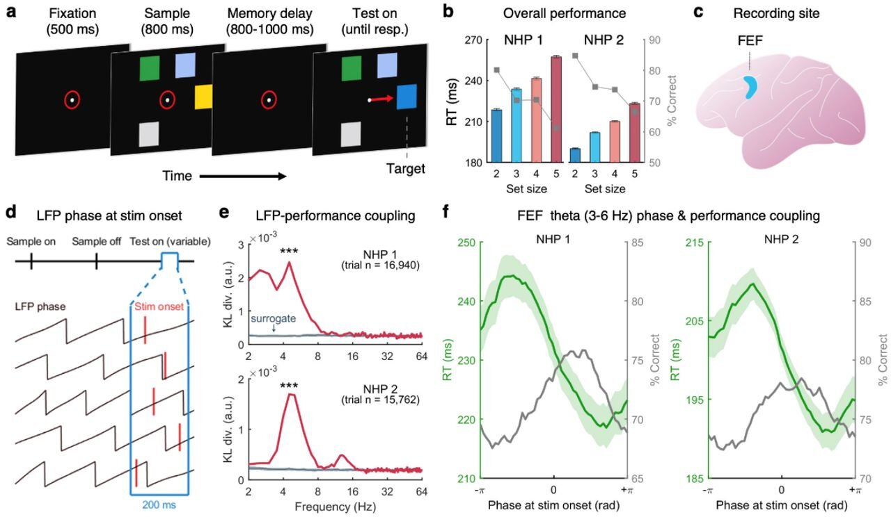
In the study, animals played a video game in which an array of colored squares appeared and then disappeared on a screen. After about a second, the array reappeared with one square sporting a different color. The animals had to glance at the one that changed, ideally as quickly as possible. To keep score, the researchers tracked the reaction time and position of the animals’ gaze.
They also measured brain wave power across a broad frequency spectrum and individual neural electrical “spikes” in a region called the “frontal eye fields” that maps visual information analogously to where it first hits the retina (a so-called “retinotopic” map).
In their post-hoc analysis of hundreds of trials of the game, the researchers noticed that the animals’ accuracy and speed turned out to depend on a combination of the phase of a theta frequency brainwave when the changed square appeared, and the vertical location on the screen of that target square.
In other words, each height on the screen had its own phase of the theta wave where performance was at its best, and the lower a target square appeared on the screen, the later the phase of the wave that correlated with peak performance.
“The optimal theta phase for behavior varied by retinotopic target location, progressing from the top to the bottom of the visual field,” the researchers wrote in Neuron. “This could be explained by a traveling wave of activity across the cortical surface during the memory delay.”
The finding suggests that the brain’s system for spotting changes in its visual field has some degree of rhythmic variability. If the wave phase at the time the changed square appeared was optimal for where the square was, performance was better.
If it wasn’t, performance was worse. Miller said his lab will need to continue studying the phenomenon to understand why it might have evolved the way it did. Theta is a commonly observed mechanism of attention when animals have to monitor more than one location at a time, Miller noted.
Further findings
Other findings in the study add to the evidence base Miller’s lab has been building for how the brain uses waves in its operations and computations. Numerous studies by his lab, for instance, have shown that alpha and beta frequency waves (8–25 Hz) impose the brain’s understanding of the rules of a task and regulate when faster gamma frequency waves (30Hz and above) can be used to encode data from the senses.
Sure enough, in this study, the lab observed that theta waves seemed to orchestrate that rivalry between beta and gamma. In the excitatory phase of the theta waves, beta was suppressed and visual information was evident in the neural spiking activity. In the inhibitory phase of theta, beta power was stronger and the spiking decreased.
The study also showed that the effect of the theta wave on performance increased depending on how many squares the animals had to remember. That raises a potential clinical implication, Miller said.
His lab is working to develop closed-loop analog feedback systems that can strengthen the power of waves at different frequencies. In disorders where theta power is too low, the study suggests that visual working memory capacity could suffer, but that intervention could help.
In addition to Han and Miller, the study’s other authors are Scott Brincat, a research scientist in Miller’s lab, and Timothy Buschman, a professor at Princeton University.
More information:
Hio-Been Han et al, Working memory readout varies with frontal theta rhythms, Neuron (2025). DOI: 10.1016/j.neuron.2025.09.031. www.cell.com/neuron/fulltext/S0896-6273(25)00744-5
Citation:
Like radar, a brain wave sweeps a cortical region to read out information held in working memory (2025, October 20)
retrieved 20 October 2025
from https://medicalxpress.com/news/2025-10-radar-brain-cortical-region-held.html
This document is subject to copyright. Apart from any fair dealing for the purpose of private study or research, no
part may be reproduced without the written permission. The content is provided for information purposes only.