Macrophages, also known as scavenger cells, are part of our immune system. They destroy invading pathogens and are found in the organs and the bloodstream. Led by Prof. Dr. Peter Wieghofer, professor of cellular neuroanatomy, researchers at the Faculty of Medicine at the University of Augsburg in Germany have now gained new insights into these immune cells in the eye.
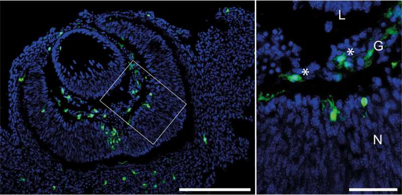
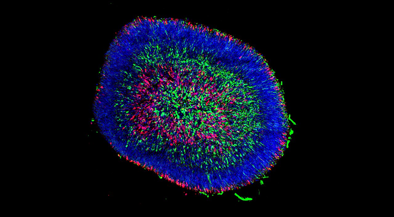
For the first time, they have shown that in the eye of the mouse macrophages are already forming in the vitreous body in the embryonic stage. For decades, the state of research was that so-called hyalocytes, which were researched at the time by Rudolf Virchows, regularly regenerate from blood cells. The new findings have been published in the Journal of Neuroinflammation.
“The macrophages in the vitreous body develop before birth, settle in the vitreous body and stay there throughout the course of life, just like the macrophages in the retina adjacent to the vitreous body, the so-called microglia,” explains Wiefhofer. As they are not, as previously assumed, renewed from cells in the blood, immunological aging processes can lead to cellular dysfunction and thus contribute to the development of diseases of the vitreous body and the retina.
“We have contributed to fundamental research,” says Wieghofer. Fundamental research concerns scientific questions that are initially concerned with gaining a better understanding of complex functions of the body in order to gain new fundamental insights.
It is not immediately known how this knowledge can be applied. However, all forms of therapy, the development of medications, and an understanding of how diseases develop are always based on the findings of fundamental research.
Hope for diabetic retinopathy
The discovery may be a starting point for the therapy of all diseases that affect the hyalocytes of the vitreous body as well as the neighboring retina, as for example with diabetic retinopathy. In the case of advanced diabetes, this retinal disease can lead to a loss of visual acuity as well as hemorrhages and damage to the retina.
“The inflammatory component of diabetic retinopathy is essentially transmitted by macrophages, which are beneficial in the early stages but harmful where there is chronic inflammation, where they can even promote vascularization, namely the formation of new blood cells.
“We hope that this harmful inflammatory reaction may someday be addressed through therapeutic approaches that act on the hyalocytes and reduce their influence on the pathological formation of new blood cells. This would reduce the danger of spontaneous hemorrhaging and reduce the risk of patient blindness,” explains Wieghofer.
Another field of research in which hyalocytes play a central role is in defects concerning the breakdown of vessels in the vitreous body during prenatal development. These defects can lead to severe hemorrhages and blindness within a very short period of time following birth. While with diabetic retinopathy, macrophages can contribute to the development of harmful blood vessels, in this context they are important for breaking down superfluous vessels.
“The new findings also offer an interesting approach for potential therapies here. The better we understand the fundamental properties of these cells, the more likely we can influence diseases at the interface of the retina and vitreous body, reducing the severity of such diseases, thus having a lasting impact on the lives of patients,” adds Wieghofer.
More information:
Dennis-Dominik Rosmus et al, Redefining the ontogeny of hyalocytes as yolk sac-derived tissue-resident macrophages of the vitreous body, Journal of Neuroinflammation (2024). DOI: 10.1186/s12974-024-03110-x
Citation:
Study shows macrophages form in eye before birth, offering hope for diabetic retinopathy treatment (2024, August 19)
retrieved 13 September 2024
from https://medicalxpress.com/news/2024-08-macrophages-eye-birth-diabetic-retinopathy.html
This document is subject to copyright. Apart from any fair dealing for the purpose of private study or research, no
part may be reproduced without the written permission. The content is provided for information purposes only.


