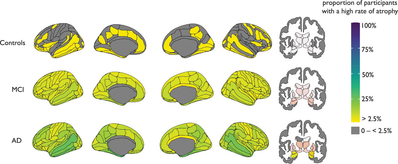
The way in which brains shrink in those who develop Alzheimer’s disease follows no specific or uniform pattern, finds a new study by researchers at UCL and Radboud University in the Netherlands.
Published in Alzheimer’s & Dementia, the study is the first to compare individual patterns of brain shrinkage over time in people with mild memory problems or Alzheimer’s disease against a healthy benchmark.
Assessing brain scans for the “fingerprints” of disease, researchers found that, among people with mild memory issues, those who develop a greater number of ‘outlier’ changes more quickly are more likely to develop Alzheimer’s. An outlier is classified as a specific brain area that, when adjusted for age and sex, has shrunk more than normal.
However, researchers also found that, despite some overlap, there was no uniform pattern to the way the brain shrank in those who developed Alzheimer’s.
The scientists say this new finding could enable more personalized medicines to be developed, targeting the specific range of brain areas affected in an individual.
Study author, Professor Jonathan Schott from UCL Queen Square Institute of Neurology, said, “We know that Alzheimer’s affects everyone differently. Understanding and quantifying this variability has important implications for the design and interpretation of clinical trials, and potentially in due course for counseling patients and developing personalized approaches to treatment.”
Alzheimer’s disease is the most common cause of dementia, accounting for 60–80% of the 944,000 people living with dementia in the UK. Previous group level studies have shown that collectively, patients with Alzheimer’s disease have excess brain shrinkage compared to healthy controls, and this can be measured using MRI scans.
However, these studies can miss how the pattern of shrinkage varies between individuals, and researchers believe this information could provide valuable information about how an individual’s cognitive performance (thinking ability/memory recall) changes over time.
To overcome this, researchers at UCL used a normative modeling approach to gain insights into individual variability between Alzheimer’s patients.
Using data from the Alzheimer’s Disease Neuroimaging Initiative (ADNI) they compared 3,233 MRI brain scans from 1,181 people with Alzheimer’s disease or mild memory issues to benchmark brain scan data collected from 58,836 healthy people. The MRI scans were typically taken at one-year intervals, with most participants having two or three scans.
Each scan was then processed using special imaging tools, that can assess the structure (thickness and volume) of the brain, across 168 different regions. This data enabled researchers to develop individualized brain maps for each participant, that could be charted over time against healthy maps (the benchmark).
The analysis showed that despite most participants starting out with similar-sized brains, different patterns (progression/regions affected) of brain shrinkage were seen between individuals over time.
Patients with Alzheimer’s disease on average had 15 to 20 outlier brain regions at the start of the study and ended up with around 30 after three years. In contrast, patients with mild memory issues started with around five to 10 outliers and accumulated only two to three more outliers over this period, on average. Importantly, having a higher number of outlier regions was associated with poorer memory in both groups.
Among people with mild memory issues, those who went on to develop dementia (within three years) accumulated four new outliers every year, while in people who remained with mild issues, the average was less than one new outlier per year.
Researchers say this new understanding could eventually help to predict how an individual’s Alzheimer’s illness will progress based on the early changes in their brain, identified in scans. However, further research is needed to pinpoint which brain changes are predictive of which future symptoms.
Professor James Cole, senior author of the study from UCL Computer Science and UCL Queen Square Institute of Neurology, said, “If we look at which areas of the brain have shrunk most in different people with Alzheimer’s, there’s no single pattern. The approach taken in our study means we can get a better sense of individual variability in Alzheimer’s disease progression. By making these brain maps, which are unique ‘fingerprints’ of a patient’s brain health, we can spot if separate brain regions are changing and how rapidly.
“While we’re still a long way from predicting exactly how an individual’s disease will progress, this approach should help us monitor the different ways people’s brains can change as symptoms develop and worsen. Hopefully, this is a step towards a more personalized approach to diagnosis and treatment.”
All people experience some form of cognitive decline as they age, and the results showed areas of overlap in brain shrinkage between healthy people and those with Alzheimer’s and /or mild memory problems. These overlap areas included the hippocampus, amygdala and other parts of the medial temporal lobe known to be critical for memory, spatial cognition, and emotion.
But the authors argue that focusing on certain areas over others may detract from the big picture.
Dr. Serena Verdi, first author of the study from UCL Computer Science and UCL Queen Square Institute of Neurology, said, “While it’s true that some regions of the brain, such as the hippocampus, are particularly important in Alzheimer’s disease, we wanted to avoid focusing on specific regions in this study. Our results confirm that everyone is different, the regions affected by disease in one person may not be the same in the next.
“I think we need to pivot towards a new way of thinking to get away from the idea that ‘this area is important, this area isn’t’. The big picture and the individual variability contained within it, is what counts.”
The authors say that some of this individual variability may stem from the fact that many people with Alzheimer’s have more than one cause of cognitive illness, such as vascular dementia or fronto-temporal dementia. Genetic and environmental factors, such as brain injuries, alcohol consumption or smoking habits, are also thought to play a part.
More information:
Serena Verdi et al, Personalizing progressive changes to brain structure in Alzheimer’s disease using normative modeling, Alzheimer’s & Dementia (2024). DOI: 10.1002/alz.14174
Citation:
Study finds no uniform brain shrinkage pattern in Alzheimer’s (2024, October 4)
retrieved 5 October 2024
from https://medicalxpress.com/news/2024-10-uniform-brain-shrinkage-pattern-alzheimer.html
This document is subject to copyright. Apart from any fair dealing for the purpose of private study or research, no
part may be reproduced without the written permission. The content is provided for information purposes only.

