Even though psychiatric disorders such as schizophrenia, bipolar disorder, and autism spectrum disorder (ASD) are quite common, their diagnosis and treatment are challenging. While doctors today have a good idea of the clinical symptoms caused by these disorders, our overall understanding of their biological characteristics and underlying physiological causes remains obscure.
Experts agree that problems with synapses—the connections that allow communication between neurons—might be a defining feature of many psychiatric disorders.
In theory, if we could analyze the characteristics of synapses in patients with psychiatric disorders, it would be possible to understand their biological basis. However, thus far, observing synapses in live humans has been challenging, and the progress in this area has been limited.
Fortunately, researchers from Yokohama City University (YCU) and other institutions in Japan have been actively working toward a solution to this problem.
In a paper published in Molecular Psychiatry on October 15, 2024, a team of researchers led by Professor Takuya Takahashi from YCU reported a new technology to visualize α-amino-3-hydroxy-5-methyl-4-isoxazole propionic acid (AMPA) receptors using positron emission tomography (PET) and a special chemical tracer called [11C]K-2.
As AMPA receptors are one of the most important molecules in neurotransmission, this method could potentially teach us a lot about the mechanisms of psychiatric disorders.
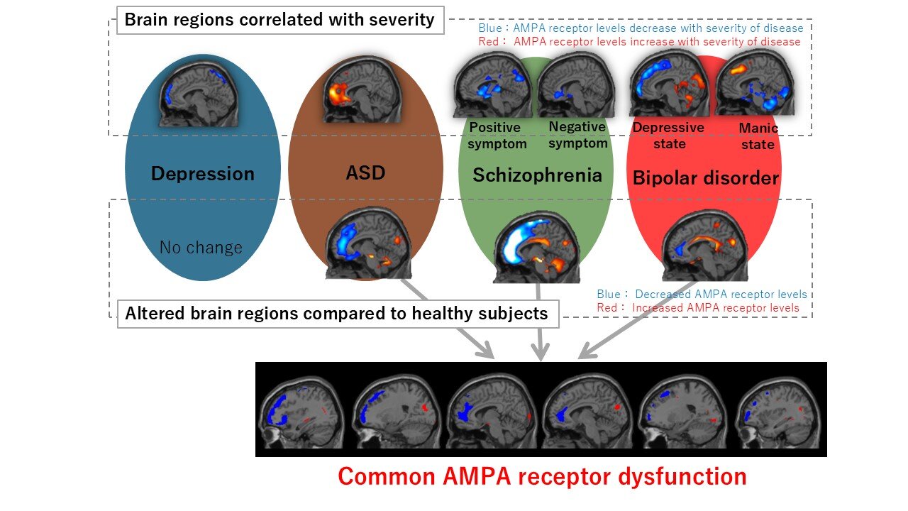
In this study, Takahashi and colleagues conducted PET scans of the brains of 149 patients with psychiatric disorders. Using the [11C]K-2 tracer, they visualized the density of AMPA receptors throughout different regions of the brain and explored its relationship with disease severity for ASD, depression, schizophrenia, and bipolar disorder.
In addition to this, the team also compared AMPA receptor density between patients with these disorders and healthy subjects.
“Identifying both unique and shared brain regions with altered AMPA receptor density offers new insights into the biological mechanisms of psychiatric disorders,” highlights Takahashi.
Their analysis suggests that an overall reduction and/or an unbalanced distribution of AMPA receptors could underlie the origin of various psychiatric disorders. It also revealed interesting characteristics of each disease. For example, in schizophrenia, the areas associated with positive symptoms (such as hallucinations) did not always overlap with areas associated with negative symptoms (such as lack of motivation and reduced emotional expression).
This suggests that the interplay between common and symptom-specific brain areas regulates the expression of different symptoms. Meanwhile, in ASD, symptom severity was largely associated with a significant increase in AMPA density throughout most of the cortex.
This elevated synaptic activity might be responsible for disrupting the process of sensory perception, which may be the cause of external sensory information overflow, often experienced by patients with ASD.
Except for depression, the other three psychiatric disorders resulted in marked differences in AMPA receptor density in various areas of the brain compared to healthy subjects. While some of these affected areas were the same across disorders, each exhibited an overall unique distribution of AMPA receptors.
Interestingly, there was almost no overlap between the regions where AMPA receptor density was correlated to disease severity (state regions) and regions that were different between healthy and diseased subjects (trait regions).
While the reason for this is not clear yet, the researchers hypothesize that trait regions may develop first and be passive indicators of a given disease, whereas the state regions may develop later and be more directly responsible for the manifestation of symptoms and their severity. Of course, this theory warrants further studies in animal models and humans alike to gather more substantial evidence.
Overall, the findings of this study showcase the power of the developed technology for visualizing important synaptic receptors and characterizing psychiatric diseases at a biological level.
“These findings could have a significant impact on the health care and pharmaceutical industries by providing a novel method for diagnosing and understanding psychiatric disorders through AMPA receptor imaging,” concludes Takahashi. “It may lead to new targeted therapies and diagnostics based on synaptic function, improving treatment precision and outcomes.”
More information:
Mai Hatano et al, Characterization of patients with major psychiatric disorders with AMPA receptor positron emission tomography, Molecular Psychiatry (2024). DOI: 10.1038/s41380-024-02785-1
Provided by
YCU Advanced Medical Research Center
Citation:
Scanning synaptic receptors: New imaging method sheds light on psychiatric disorders (2024, November 5)
retrieved 5 November 2024
from https://medicalxpress.com/news/2024-11-scanning-synaptic-receptors-imaging-method.html
This document is subject to copyright. Apart from any fair dealing for the purpose of private study or research, no
part may be reproduced without the written permission. The content is provided for information purposes only.


