A recent position paper in the Asia-Pacific Journal of Ophthalmology explores the transformative potential of artificial intelligence (AI) in ophthalmology.
With fundus photography enabling the visualization of retina at the back of the eye, the potential of AI in providing systemic disease biomarkers is becoming a reality. When fundus images are of sufficient quantity and quality, it becomes possible to train AI systems to detect elevated HbA1c levels—an important marker for high blood sugar that is traditionally obtained with blood draws, which indicates a heightened risk of diabetes and cardiovascular disease. This process leverages the emerging field of oculomics, which studies ophthalmic biomarkers to gain insights into systemic health.
In their manuscript, titled “Development of Oculomics Artificial Intelligence for Cardiovascular Risk Factors: A Case Study in Fundus Oculomics for HbA1c Assessment and Clinically Relevant Considerations for Clinicians,” this multi-institutional team explores the potential of oculomics and highlights pertinent topics for clinicians to consider as we move into an era where artificial intelligence has the potential to enhance systemic health through eye care.
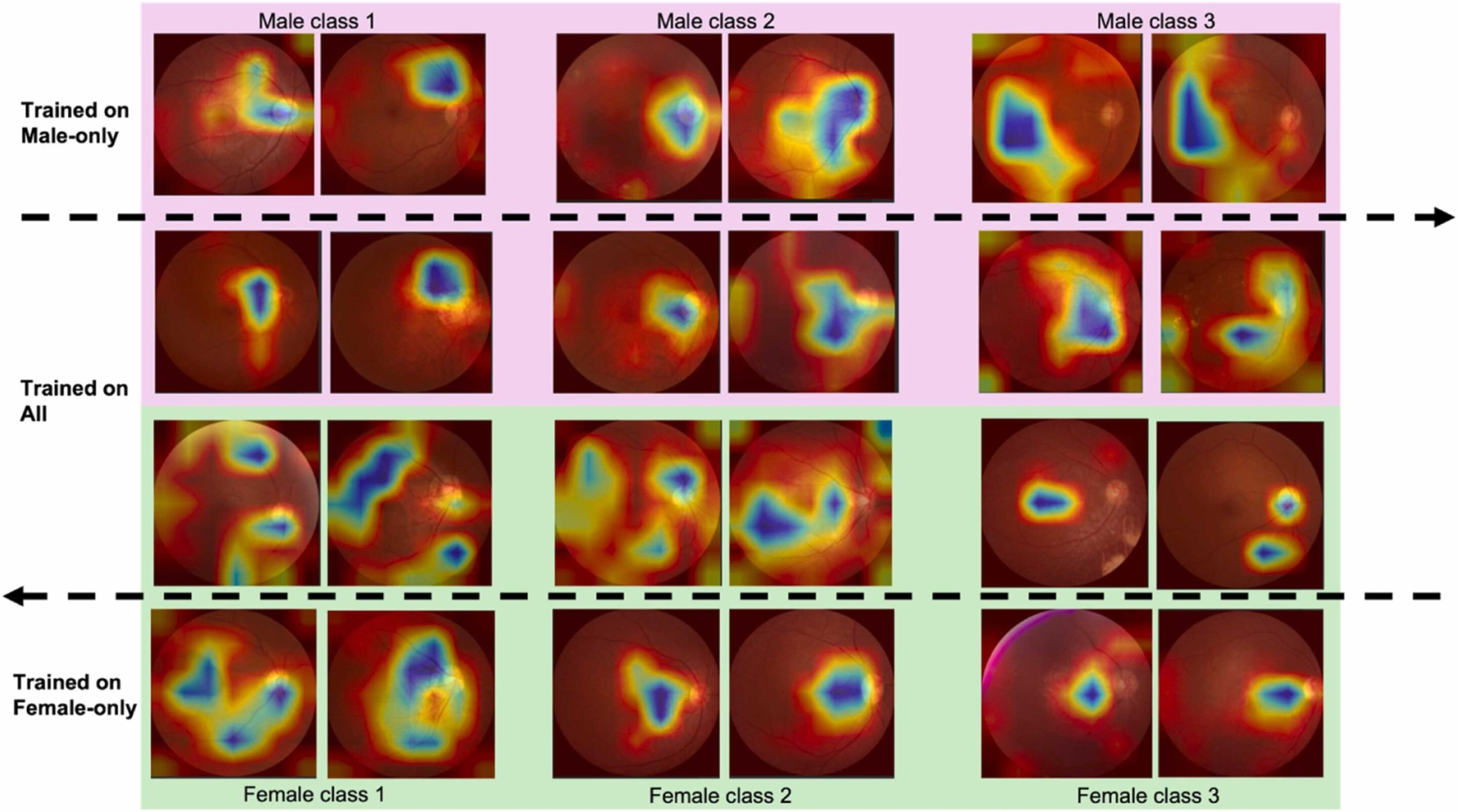
Their discussion is supported by preliminary research results from a pilot study that trained AI models to predict HbA1c levels based on fundus images. This study evaluated various factors—such as AI model size and architecture, the presence of diabetes, and patient demographics (age and sex)—and their impact on AI performance.
One of the study observations was that biased training samples for an oculomics model, such as a pool of predominantly older patients, can degrade model performance. The results of the case study highlight the importance of developing trustworthy AI models for assessing cardiovascular risk factors while addressing the challenges and problems that must be overcome prior to clinical adoption, as well as advancing reliable oculomics technology.
“By leveraging AI to analyze retinal images for cardiovascular risk assessment,” says Al-Aswad, “we aim to bridge a crucial gap in early disease detection.
“This method not only enhances our ability to identify at-risk individuals but also holds promise for transforming how we manage chronic conditions such as diabetes. By focusing on practical applications of this technology, we are advancing towards more personalized and preventative health care solutions.”
“While these advancements hold promise, it is also of utmost importance for clinicians and researchers to develop and employ these techniques in a responsible manner, as this will benefit patient care the most in the end,” adds Kuk Jin Jang, a postdoctoral researcher in the Penn Research in Embedded Computing and Integrated Systems Engineering (PRECISE) Center at the University of Pennsylvania.
“Our collaboration serves to further understand how we can responsibly leverage this revolutionary technology to benefit patients in the future. It is a testament to the collaborative advances formed when health care and engineering come together to work towards responsible AI for patient care,” says Joshua Ong, a resident physician at the University of Michigan and PRECISE Center affiliate.
“I am incredibly grateful for our multidisciplinary team for coming together to bring this paper and topic to the forefront.”
“This collaboration reflects a deep commitment to advancing health care through innovative AI applications,” adds PRECISE Center Director Insup Lee, Cecilia Fitler Moore Professor in Computer and Information Science at Penn Engineering.
“By combining our expertise, we are paving the way for significant improvements in patient care and the overall management of long-term health challenges.”
Led by Lama Al-Aswad, Professor of Ophthalmology and Irene Heinz Given and John La Porte Given Research Professor of Ophthalmology II, of the Scheie Eye Institute, the work represents a collaboration among researchers from Penn Engineering, Penn Medicine, the University of Michigan Kellogg Eye Center, St. John Eye Hospital in Jerusalem, and Gyeongsang National University College of Medicine in Korea.
More information:
Joshua Ong et al, Development of oculomics artificial intelligence for cardiovascular risk factors: A case study in fundus oculomics for HbA1c assessment and clinically relevant considerations for clinicians, Asia-Pacific Journal of Ophthalmology (2024). DOI: 10.1016/j.apjo.2024.100095
Citation:
Leveraging AI to analyze retinal images for cardiovascular risk assessment (2024, October 4)
retrieved 5 October 2024
from https://medicalxpress.com/news/2024-10-leveraging-ai-retinal-images-cardiovascular.html
This document is subject to copyright. Apart from any fair dealing for the purpose of private study or research, no
part may be reproduced without the written permission. The content is provided for information purposes only.


