Important insights into the mechanisms behind Fuchs endothelial corneal dystrophy (FECD), a common cause of age-related visual loss, have been revealed in a new study led by UCL researchers.
The study, published in eBioMedicine, provides hope for future therapeutic developments, and finds implications for other neurological diseases.
FECD is a common, inherited eye condition that primarily affects the cornea, the clear front part of the eye. It is one of the leading causes of vision loss as people age and is the most common reason for corneal transplants in high-income countries. FECD affects the corneal endothelial cells, which form a layer responsible for controlling fluid balance in the cornea. When these cells are lost more quickly than usual in people with FECD, the cornea becomes swollen and cloudy, leading to blurred vision.
The research, led by a team in Dr. Alice Davidson’s Inherited Corneal Disease Lab at UCL Institute of Ophthalmology, has revealed how FECD progresses at a molecular level, and highlights the importance of understanding genetic instability—when cells have high frequency of mutations—in developing new treatments for FECD and diseases caused by similar genetic mutations, such as Huntington’s disease and other neurological and neuromuscular diseases.
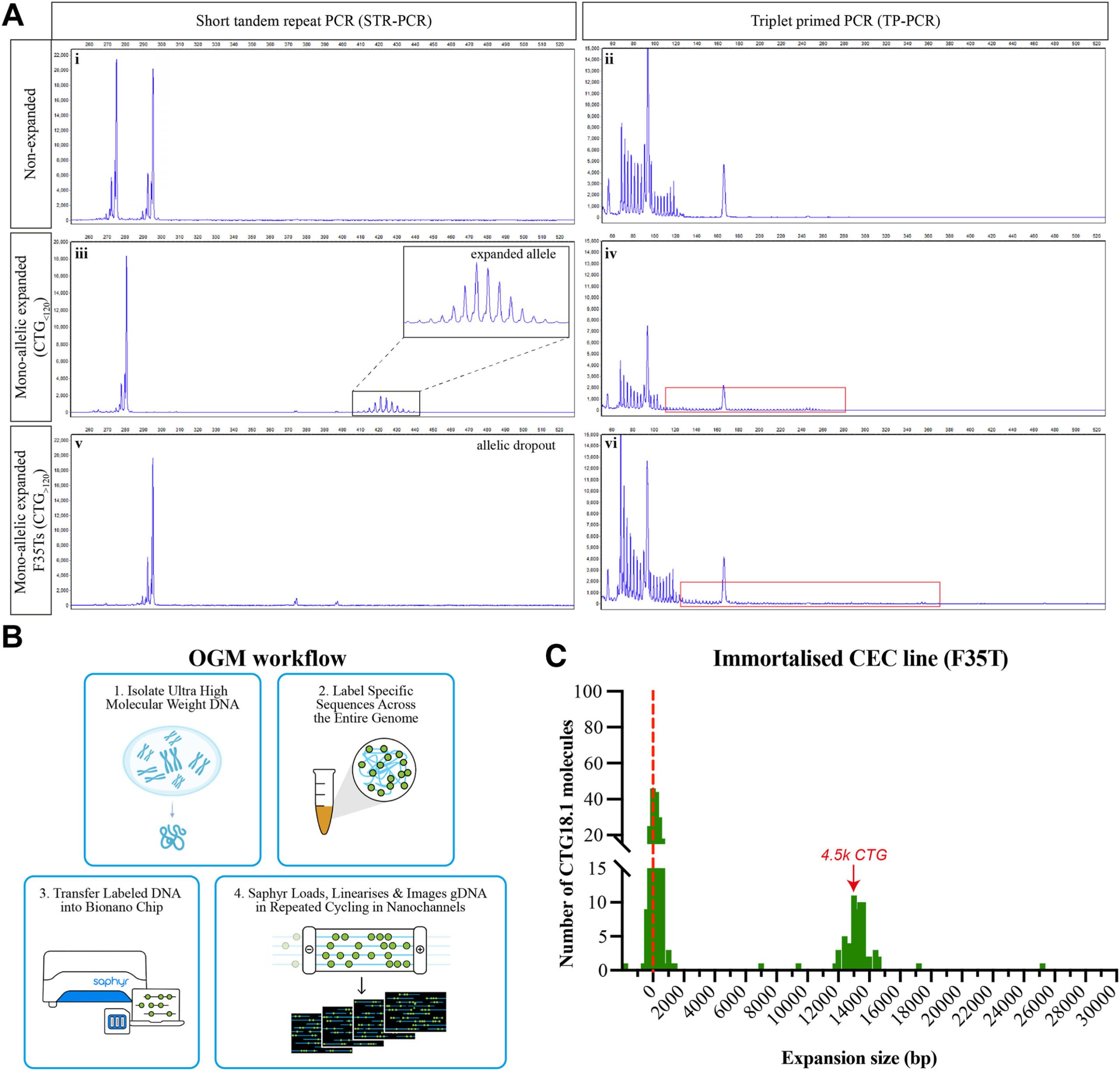
The study utilized advanced optical genome mapping with single-molecule precision where researchers found extreme levels of instability to identify how the disease progresses. These findings were exclusively in the corneal endothelial cells of individuals with FECD. The study also identified that both size and patient age influence instability rates.
Dr. Christina Zarouchlioti (UCL Institute of Ophthalmology), lead author, said, “We are excited to share these results and the impact they might have for the future of patients with FECD. We also know that the study’s implications extend beyond FECD, positioning it as a valuable model for understanding a growing number of other diseases, such as Huntington’s disease and myotonic dystrophies, which share similar mechanisms.”
A key factor in developing FECD is the expansion of a specific DNA sequence within the TCF4 gene, called CTG18.1. This genetic change, known as a short tandem repeat expansion, has been identified as the most common risk factor for FECD across all studied populations.
Researchers are now focused on a new series of experiments to understand how this mechanism plays out throughout human development to better understand when may be the most effective time to therapeutically intervene.
More information:
Christina Zarouchlioti et al, Tissue-specific TCF4 triplet repeat instability revealed by optical genome mapping, eBioMedicine (2024). DOI: 10.1016/j.ebiom.2024.105328
Citation:
Key mechanism behind common genetic cause of age-related visual loss discovered (2024, September 19)
retrieved 20 September 2024
from https://medicalxpress.com/news/2024-09-key-mechanism-common-genetic-age.html
This document is subject to copyright. Apart from any fair dealing for the purpose of private study or research, no
part may be reproduced without the written permission. The content is provided for information purposes only.


