
Age-related macular (AMD) degeneration is a leading cause of vision impairment and blindness in the elderly population. In so-called wet AMD, new, abnormal blood vessels grow in the central part of the retina called the macula, which is required for high-acuity central vision, leading to fluid and blood leakage and macular damage or dysfunction. Although wet AMD accounts for a minority of AMD cases, 90% of AMD-related cases of blindness are due to wet AMD.
Wet AMD in its early stages can be treated with drugs to reduce the formation of new blood vessels, but this treatment is inefficient in cases where blood vessel formation is already in its advanced stages.
A new alternative treatment for those patients may be surgical removal of the abnormal blood vessels followed by the transplantation of stem cell-derived retinal cells, according to a study led by Yong Liu and colleagues from Third Military Medical University Southwest Hospital, China, published in the journal Stem Cell Reports.
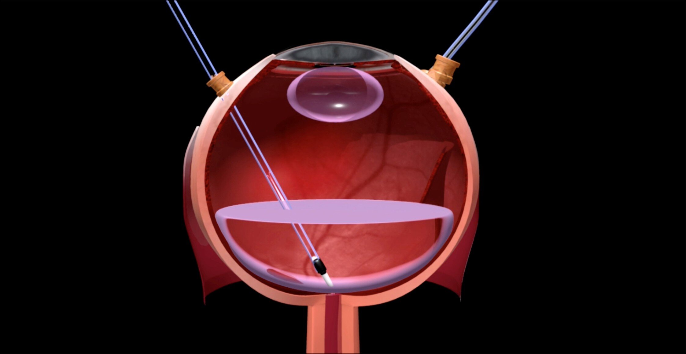
In their clinical study, involving 10 patients with wet AMD, the researchers first developed a method for safely removing the newly formed blood vessels followed by the transplantation of stem cell-derived retinal cells to replace the patients’ damaged or lost retinal cells.
The retinal structure improved in those patients where blood vessel patches were completely removed during surgery, suggesting that the transplanted cells survived and repaired the damaged retina. Further, visual acuity remained stable or improved in those patients during the 12-months follow-up, with limited side effects.
In contrast, patients where the blood vessel patches could only partially be removed experienced persistent bleeding and inflammation in the eye and an incomplete regeneration of the retina, and vision did not improve in those patients.
The researchers concluded that complete and safe removal of the blood vessel patches prevents inflammation and generates a milieu favorable for transplant survival and integration.
Follow-up studies with larger groups of patients are required to confirm the clinical efficacy and favorable safety profile of this type of treatment.
More information:
Safe CNV removal is crucial for successful hESC-RPE transplantation in wet age-related macular degeneration, Stem Cell Reports (2025). DOI: 10.1016/j.stemcr.2025.102424. www.cell.com/stem-cell-reports … 2213-6711(25)00028-1
Provided by
International Society for Stem Cell Research
Citation:
Stem cell transplant clears clinical safety hurdle for treatment of wet age-related macular degeneration (2025, February 27)
retrieved 27 February 2025
from https://medicalxpress.com/news/2025-02-stem-cell-transplant-clinical-safety.html
This document is subject to copyright. Apart from any fair dealing for the purpose of private study or research, no
part may be reproduced without the written permission. The content is provided for information purposes only.