
A new method of taking microscopic images of a live mouse’s retina through the eye allows researchers to record the reaction of brain cells to disease and treatment. The Kobe University development is more easily applicable than previous methods and promises to advance research on and treatment of vision-related diseases.
Diabetic retinopathy, a form of diabetic eye disease, is one of the leading causes of blindness around the globe. “It’s understood that vision is lost due to damage to the blood vessels in the retina, but recent research has identified that abnormalities in neurons and immune cells begin prior to vascular damage,” says Kobe University neurophysiologist Tachibana Yoshihisa.
He continues, “In particular, microglia, immune cells that reside in the retina and constantly monitor their environment, initiate inflammation when abnormalities occur. But because it is difficult to observe their behavior in living organisms, much of their involvement has remained in the dark.”
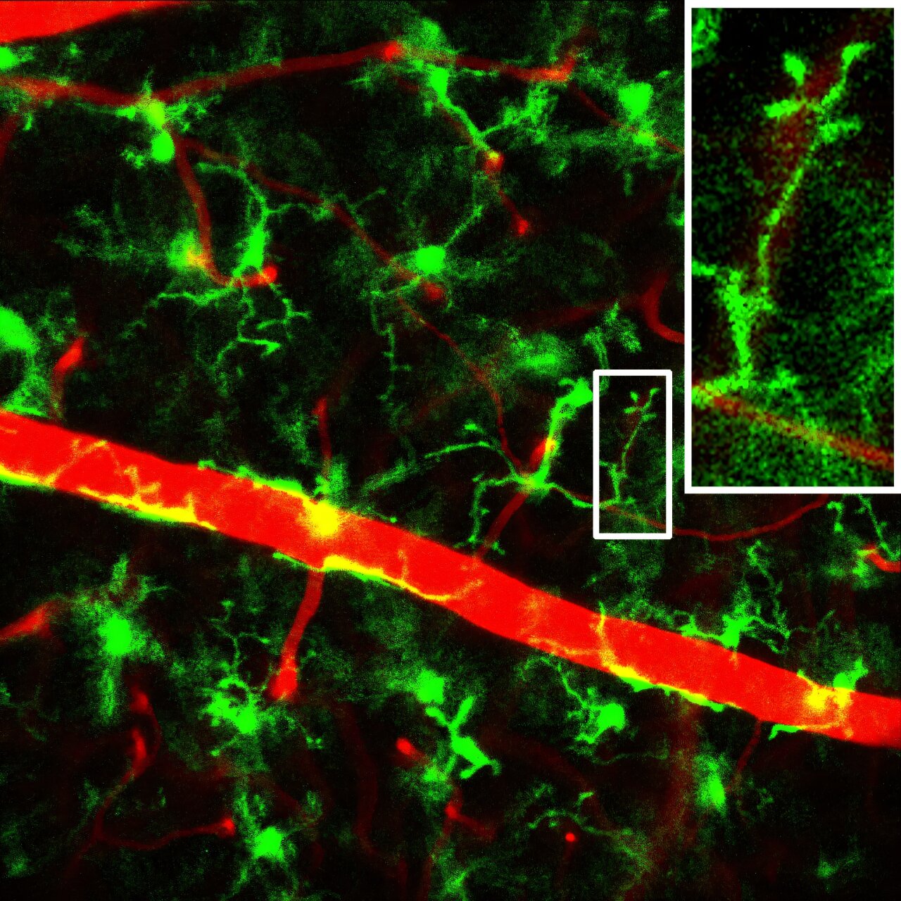
Conventional microscopy setups either require advanced technical expertise to correct the distorted images or don’t achieve high-resolution live images with readily available technology. That’s why Tachibana and his team developed a new technology that combines a head-fixation device, custom-made contact lenses and a special but commercially available objective lens.
“This approach allows us clear, long-term observation of the living retina, down to the minute movements of microglia,” the Kobe University researcher says.

In the journal PNAS, Tachibana and his team report that their newly developed method allowed them to identify that microglia start to move more actively in diabetic mice, indicating increased monitoring activity, long before tissue damage is noticeable.
“This phenomenon has been overlooked in conventional observation in non-living specimens and is an important finding that provides a new perspective for understanding the pathology of diabetic retinopathy,” explains Tachibana.
His team also observed the effect of the diabetes drug liraglutide on microglia. They found that in diabetic mice, the heightened activity of microglia was returned to normal, but also that the activity of these cells got reduced in healthy mice, too. In addition, the drug did not alter sugar levels in the blood.
Tachibana says, “This suggests that liraglutide acts on microglia through a mechanism that directly modulates their behavior.”
With insights like these becoming possible, the ability to observe the behavior of cells in the living organism directly is of great advantage for developing new treatments.
“We expect this technology to be useful for other retinal diseases such as glaucoma and age-related macular degeneration, too,” says Tachibana. But the Kobe University researcher also has his sight on another goal.
He says, “Blindness by diabetic eye disease is a serious global challenge. We hope that our technology may be put to practical use in clinical settings, as a non-invasive diagnostic method, making the eye a window to spotting systemic disease.”
More information:
Tachibana, Yoshihisa et al, Transpupillary in vivo two-photon imaging reveals enhanced surveillance of retinal microglia in diabetic mice, Proceedings of the National Academy of Sciences (2025). DOI: 10.1073/pnas.2426241122. doi.org/10.1073/pnas.2426241122
Citation:
Live retinal imaging in mice reveals early immune cell activity before diabetes-related damage occurs (2025, October 6)
retrieved 6 October 2025
from https://medicalxpress.com/news/2025-10-retinal-imaging-mice-reveals-early.html
This document is subject to copyright. Apart from any fair dealing for the purpose of private study or research, no
part may be reproduced without the written permission. The content is provided for information purposes only.