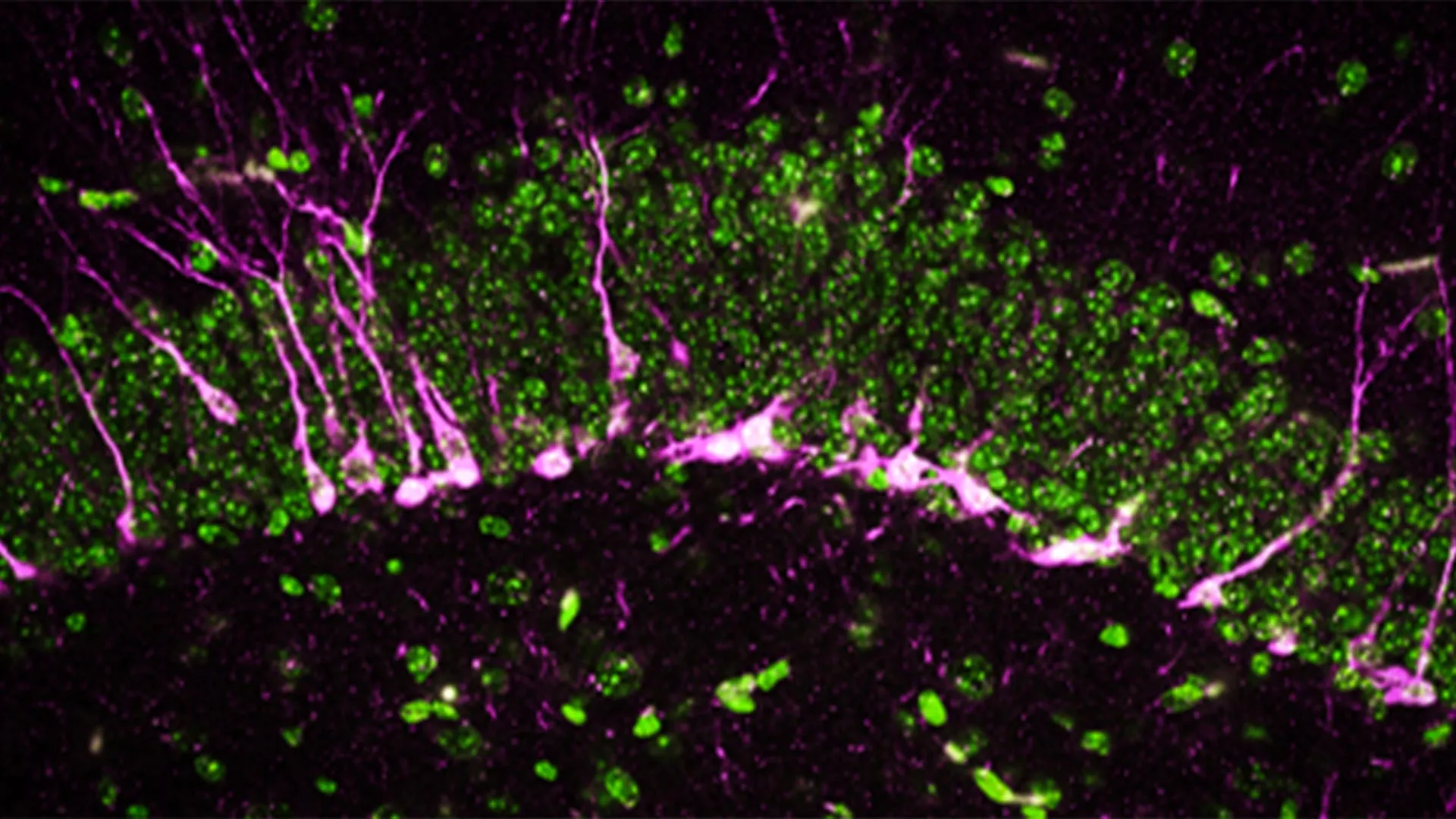
Researchers are investigating the mechanisms and identifying new areas of the brain that can benefit patients when stimulated.
Persons with Parkinson’s disease increasingly lose their mobility over time and are eventually unable to walk. Hope for these patients rests on deep brain stimulation, also known as a brain pacemaker. In a current study, researchers at Ruhr University Bochum and Philipps-Universität Marburg, Germany, investigated whether and how stimulation of a certain region of the brain can have a positive impact on ambulatory ability and provide patients with higher quality of life. To do this, the researchers used a technique in which the nerve cells are activated and deactivated via light. Their report appeared in the journal Scientific Reports on April 12, 2025.
Improving ambulatory ability
If medication is no longer sufficient in alleviating restricted mobility in the advanced stage of Parkinson’s disease, one alternative is deep brain stimulation. An electrical pulse emitter is implanted within the brain, such as in the subthalamic nucleus, which is functionally part of the basal ganglia system.
The group under Dr. Liana Melo-Thomas from Philipps-Universität Marburg was able to show in previous studies on rats that stimulation of the inferior colliculus — chiefly known for processing auditory input — can be used to overcome mobility restrictions. “There are indications that stimulation of this region of the brain leads to activation of the mesencephalic locomotor region, or MLR,” says Melo-Thomas.
Interestingly, the colliculus inferior — unlike the basal ganglia — is not affected by Parkinson’s disease. However, the research group under Melo-Thomas discovered that its stimulation activates alternative motor pathways and can improve patients’ mobility.
The current study aimed to further investigate this activating influence of the inferior colliculus on the MLR. “We suspected that this would have a positive effect on ambulatory ability,” says Melo-Thomas.
Optically influencing nerve cells
The Marburg group led by Professor Rainer Schwarting sought support by Dr. Wolfgang Kruse from the Department of General Zoology and Neurobiology at Ruhr University Bochum. The team in Bochum led by Professor Stefan Herlitze played a significant role in co-developing the methods of optogenetics.
While doing so, the researchers ensure that the nerve cells of genetically modified test animals produce a light-sensitive protein in interesting regions of the brain. Light that reaches these nerve cells via small, implanted optical fibers allows the researchers to activate or inhibit them specifically. “This method is thus much more precise than electrical stimulation, which always affects the area around the cells as well,” says Kruse.
For the first time, the effect of the stimulation was directly documented with electrophysiological measurements of neuronal activity in the target structures. A multi-electrode system originally developed at Philipps-Universität Marburg was used for this purpose. By combining these methods, the researchers were able to directly understand the effect of the stimulation. Parallel measurement with up to four electrodes is also highly efficient, allowing minimization of the number of animals used. Behavioral effects that can be triggered by the stimulation were monitored in conscious animals.
Stimulation of the inferior colliculus provides the desired effect
Optogenetic stimulation in the inferior colliculus predominantly triggered the expected increase in neuronal activity within it. “Simultaneous measurements in the deeper MLR region showed increased activity in the majority of cells, although nearly one quarter of the cells were inhibited by the additional activity in the inferior colliculus,” reports Kruse. The activation of individual nerve cells occurred with an average delay of 4.7 milliseconds, indicating a functional synaptic interconnection between the inferior colliculus and MLR.
Foundations for new types of therapy
Investigating circuits outside of the basal ganglia that are affected by Parkinson’s disease is a promising step in the search for a new therapeutic approach to alleviating motor deficits resulting from the disease. Such is the case with the connection between the inferior colliculus and the MLR that was investigated for this study.
“Even if the path toward new therapeutic approaches to alleviating the symptoms of Parkinson’s disease still appears long, such foundational research is immensely important,” emphasizes Kruse. The exact mechanisms that lead to the observed relief of symptoms with deep brain stimulation in the basal ganglia are not fully understood. Further investigation of the underlying interconnections may provide new insight that could optimize therapy in the long term.